Placenta evaluation trial seeks to establish opposed being pregnant end result threat | Medical analysis
[ad_1]
Scientists are launching a trial of a brand new scanning approach that might establish girls at excessive threat of opposed being pregnant outcomes, together with stillbirth and pre-eclampsia, by analysing their placentas.
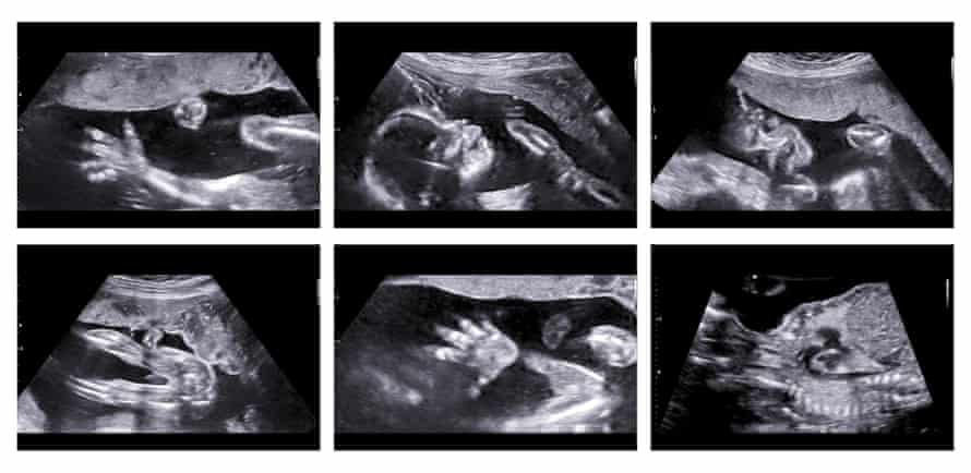
The expertise makes use of synthetic intelligence to analyse ultrasound pictures taken throughout girls’s 12-week scan and assign them a threat rating – just like the primary trimester threat evaluation for Down’s syndrome routinely provided at this level in girls’s pregnancies. These deemed at excessive threat could possibly be provided further scans or medicine to scale back their threat of opposed outcomes.
Every day, roughly eight households within the UK are affected by stillbirth, with foetal progress restriction – a situation the place the child is smaller than anticipated, or its progress slows or stops throughout being pregnant – the only greatest threat issue.
In the meantime, roughly 6% of pregnant girls develop pre-eclampsia – a situation that causes hypertension or issues with the liver or kidneys – which might be severe if untreated.
Each situations are regarded as associated to issues with the placenta, the jelly-like organ linking the foetus’s blood provide to the mom’s.

Some hospitals inside the NHS are starting to introduce a screening algorithm that makes use of routinely collected antenatal knowledge, together with an ultrasound measure of uterine blood provide, to calculate girls’s threat of pre-eclampsia, which may allow medical doctors to intervene with further screening, or early supply of the child the place crucial.
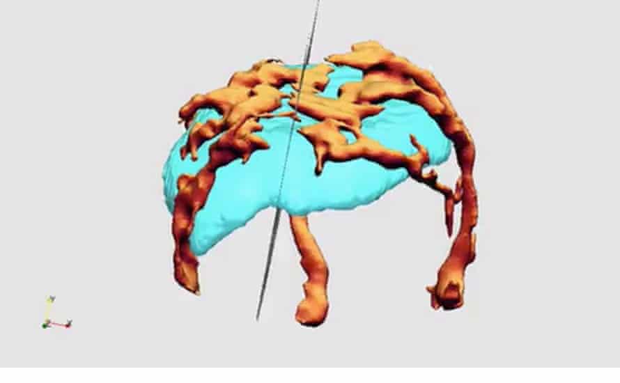
The brand new strategy goals to construct on this algorithm, by incorporating further AI-based assessments of the placenta’s quantity, and the blood vessels supplying it.
“Many research have proven that when you have a small placenta within the first trimester, you should have a small placenta at time period, and a small placenta makes a small child,” stated Prof Sally Collins, a marketing consultant obstetrician and medical lead for girls’s well being at Perspectum Ltd, an Oxford College spin-out firm that’s growing the expertise.
“In case your placenta is small, and has a standard provide of blood vessels (vascularity), it may be a sign of foetal progress restriction. Whether it is small with low vascularity, you doubtlessly get a small child and preeclampsia.”

Though the placenta might be visualised utilizing ultrasound, measuring it, and all of the tiny blood vessels supplying it, is extraordinarily time consuming, making this impractical for routine early being pregnant screening. So the College of Oxford has used machine studying to develop a device, educated on hundreds of ultrasound pictures the place the placenta has been painstakingly marked out by hand, to automate the popularity course of.
“What we’ve give you is a totally automated, synthetic intelligence technique for seeing and measuring the placenta, and the blood provide at its interface with the uterus, so all of the sudden, we have now a pc device that may inform you the dimensions and the vascularity of the placenta in actual time,” Collins stated.
A pilot examine in 143 girls recommended the device may differentiate between these with pre-eclampsia and foetal progress restriction. The brand new trial, launched this week , will check whether or not integrating this data into the prevailing NHS algorithm can additional enhance its accuracy.
It goals to recruit 4,000 girls who shall be provided a placental ultrasound at their 12-week appointment, alongside routine care, after which adopted as much as see if the improved algorithm’s predictions are appropriate.

